CT Scans (Computed Tomography scans) Applications, Benefits & Risk
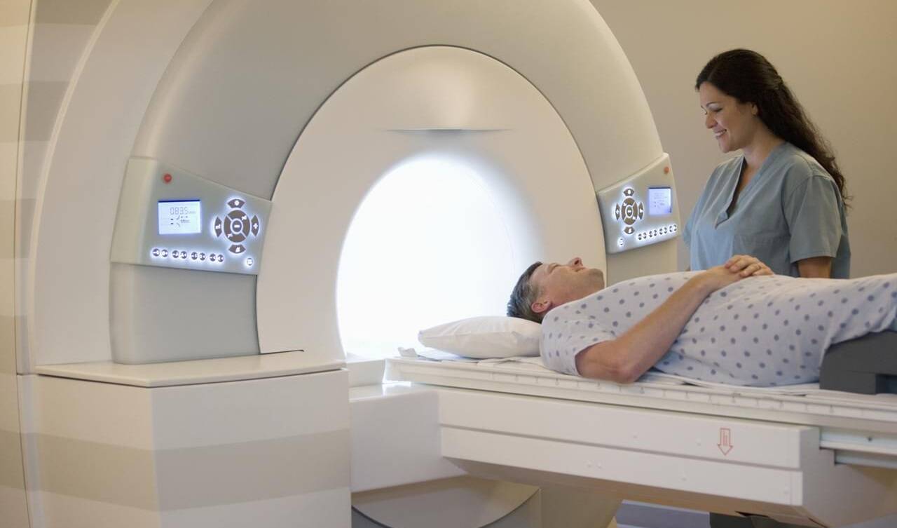
Computed Tomography (CT) scans, also known as CT imaging or CAT scans (Computed Axial Tomography), are medical imaging techniques used to create detailed cross-sectional images of various parts of the body. This powerful diagnostic tool provides valuable information to healthcare professionals for diagnosing and managing a wide range of medical conditions. In this comprehensive overview, we will explore the principles of CT imaging, its applications, benefits, risks, and advancements in technology.
Principles of CT Imaging:
CT imaging utilizes X-rays to produce detailed images of the body’s internal structures. The basic principles of CT imaging include:
- X-ray Generation:
- X-ray Absorption:
- As X-rays pass through the body, they are absorbed to varying degrees by different tissues. Dense tissues, such as bone, absorb more X-rays and appear white on the resulting images (high attenuation), while less dense tissues, such as air-filled spaces, absorb fewer X-rays and appear darker (low attenuation).
- Rotational Scanning:
- During a CT scan, the X-ray tube and detector array rotate around the patient, capturing multiple X-ray images from different angles.
- Data Acquisition:
- The X-ray attenuation data collected from multiple angles are processed by a computer to reconstruct cross-sectional images (slices) of the body.
- Image Reconstruction:
- Advanced algorithms are used to reconstruct the collected data into detailed cross-sectional images, which can be viewed and analyzed by radiologists and other healthcare professionals.
Applications of CT Scans:
CT scans are versatile imaging tools used in various medical specialties for diagnostic and treatment purposes. Some common applications include:
- Trauma Evaluation:
- CT imaging is invaluable in assessing injuries sustained in traumatic incidents, providing detailed information about fractures, internal bleeding, and organ damage.
- Diagnosis of Medical Conditions:
- CT scans are used to diagnose a wide range of medical conditions, including cancers, neurological disorders, cardiovascular diseases, and pulmonary conditions.
- Cancer Detection and Staging:
- CT scans are frequently used to detect and stage cancers, providing detailed images of tumors and their surrounding tissues.
- Evaluation of Abdominal and Pelvic Organs:
- CT imaging is commonly used to evaluate the liver, pancreas, kidneys, bladder, and other abdominal and pelvic organs for conditions such as infections, tumors, and inflammatory diseases.
- Evaluation of Brain and Spinal Cord:
- CT scans of the brain and spine can detect abnormalities such as tumors, hemorrhages, strokes, and traumatic injuries.
- Guidance for Minimally Invasive Procedures:
- CT scans provide precise guidance for minimally invasive procedures such as biopsies, drainage procedures, and catheter placements.
- Evaluation of Musculoskeletal Disorders:
- CT imaging is valuable for assessing musculoskeletal disorders, including bone fractures, joint abnormalities, and degenerative diseases.
Benefits of CT Scans:
CT scans offer several advantages over other imaging modalities, making them indispensable tools in modern healthcare:
- Speed and Efficiency:
- CT scans are relatively quick to perform, providing detailed images in a matter of minutes, making them ideal for emergency situations.
- High Resolution and Contrast:
- CT imaging produces high-resolution images with excellent tissue contrast, allowing for detailed visualization of anatomical structures and abnormalities.
- Versatility:
- CT scans can image virtually any part of the body, making them suitable for diagnosing a wide range of medical conditions.
- Non-invasive:
- CT scans are non-invasive procedures that do not require incisions or injections, reducing the risk of complications compared to invasive diagnostic techniques.
- Painless:
- Patients undergoing CT scans typically experience minimal discomfort, as the procedure is painless and requires no special preparation.
- Accessibility:
- CT scanners are widely available in hospitals and medical centers, ensuring timely access to diagnostic imaging services for patients.
Risks and Considerations:
While CT scans are valuable diagnostic tools, they are associated with certain risks and considerations:
- Radiation Exposure:
- CT scans involve exposure to ionizing radiation, which carries a small risk of potentially harmful effects, particularly with repeated or unnecessary scans.
- Contrast Agents:
- Some CT scans may require the administration of contrast agents (iodine-based or gadolinium-based) to enhance image quality. Allergic reactions and kidney damage are potential risks associated with contrast agents.
- Pregnancy Considerations:
- Pregnant women are generally advised to avoid unnecessary radiation exposure, as it may pose risks to the developing fetus. However, in certain cases, the benefits of a CT scan may outweigh the potential risks.
- Contrast-induced Nephropathy:
- Patients with pre-existing kidney disease are at increased risk of developing contrast-induced nephropathy, a condition characterized by kidney damage following contrast agent administration.
- Overutilization:
- Overutilization of CT scans, particularly in asymptomatic individuals, can lead to unnecessary radiation exposure and healthcare costs.
Advancements in CT Technology:
Advancements in CT technology continue to improve imaging quality, reduce radiation exposure, and enhance patient comfort:
- Low-Dose CT Protocols:
- Low-dose CT protocols employ techniques to minimize radiation exposure while maintaining image quality, making them suitable for certain diagnostic applications.
- Iterative Reconstruction Algorithms:
- Iterative reconstruction algorithms improve image quality and reduce noise, allowing for lower radiation doses without compromising diagnostic accuracy.
- Dual-Energy CT:
- Dual-energy CT techniques provide additional information about tissue composition and enable enhanced characterization of lesions, improving diagnostic accuracy.
- CT Angiography:
- CT angiography (CTA) is a specialized technique used to visualize blood vessels and assess blood flow, aiding in the diagnosis of vascular diseases and guiding interventions.
- Cone Beam CT:
- Cone beam CT technology is used in specialized applications, such as dental imaging and image-guided radiation therapy, offering high-resolution 3D imaging with reduced radiation exposure.
Share this article

Computed Tomography (CT) scans, also known as CT imaging or CAT scans (Computed Axial Tomography), are medical imaging techniques used to create detailed cross-sectional images of various parts of the body. This powerful diagnostic tool provides valuable information to healthcare professionals for diagnosing and managing a wide range of medical conditions. In this comprehensive overview, we will explore the principles of CT imaging, its applications, benefits, risks, and advancements in technology.
Principles of CT Imaging:
CT imaging utilizes X-rays to produce detailed images of the body’s internal structures. The basic principles of CT imaging include:
- X-ray Generation:
- CT scanners generate X-rays using an X-ray tube positioned opposite a detector array.
- X-ray Absorption:
- As X-rays pass through the body, they are absorbed to varying degrees by different tissues. Dense tissues, such as bone, absorb more X-rays and appear white on the resulting images (high attenuation), while less dense tissues, such as air-filled spaces, absorb fewer X-rays and appear darker (low attenuation).
- Rotational Scanning:
- During a CT scan, the X-ray tube and detector array rotate around the patient, capturing multiple X-ray images from different angles.
- Data Acquisition:
- The X-ray attenuation data collected from multiple angles are processed by a computer to reconstruct cross-sectional images (slices) of the body.
- Image Reconstruction:
- Advanced algorithms are used to reconstruct the collected data into detailed cross-sectional images, which can be viewed and analyzed by radiologists and other healthcare professionals.
Applications of CT Scans:
CT scans are versatile imaging tools used in various medical specialties for diagnostic and treatment purposes. Some common applications include:
- Trauma Evaluation:
- CT imaging is invaluable in assessing injuries sustained in traumatic incidents, providing detailed information about fractures, internal bleeding, and organ damage.
- Diagnosis of Medical Conditions:
- CT scans are used to diagnose a wide range of medical conditions, including cancers, neurological disorders, cardiovascular diseases, and pulmonary conditions.
- Cancer Detection and Staging:
- CT scans are frequently used to detect and stage cancers, providing detailed images of tumors and their surrounding tissues.
- Evaluation of Abdominal and Pelvic Organs:
- CT imaging is commonly used to evaluate the liver, pancreas, kidneys, bladder, and other abdominal and pelvic organs for conditions such as infections, tumors, and inflammatory diseases.
- Evaluation of Brain and Spinal Cord:
- CT scans of the brain and spine can detect abnormalities such as tumors, hemorrhages, strokes, and traumatic injuries.
- Guidance for Minimally Invasive Procedures:
- CT scans provide precise guidance for minimally invasive procedures such as biopsies, drainage procedures, and catheter placements.
- Evaluation of Musculoskeletal Disorders:
- CT imaging is valuable for assessing musculoskeletal disorders, including bone fractures, joint abnormalities, and degenerative diseases.
Benefits of CT Scans:
CT scans offer several advantages over other imaging modalities, making them indispensable tools in modern healthcare:
- Speed and Efficiency:
- CT scans are relatively quick to perform, providing detailed images in a matter of minutes, making them ideal for emergency situations.
- High Resolution and Contrast:
- CT imaging produces high-resolution images with excellent tissue contrast, allowing for detailed visualization of anatomical structures and abnormalities.
- Versatility:
- CT scans can image virtually any part of the body, making them suitable for diagnosing a wide range of medical conditions.
- Non-invasive:
- CT scans are non-invasive procedures that do not require incisions or injections, reducing the risk of complications compared to invasive diagnostic techniques.
- Painless:
- Patients undergoing CT scans typically experience minimal discomfort, as the procedure is painless and requires no special preparation.
- Accessibility:
- CT scanners are widely available in hospitals and medical centers, ensuring timely access to diagnostic imaging services for patients.
Risks and Considerations:
While CT scans are valuable diagnostic tools, they are associated with certain risks and considerations:
- Radiation Exposure:
- CT scans involve exposure to ionizing radiation, which carries a small risk of potentially harmful effects, particularly with repeated or unnecessary scans.
- Contrast Agents:
- Some CT scans may require the administration of contrast agents (iodine-based or gadolinium-based) to enhance image quality. Allergic reactions and kidney damage are potential risks associated with contrast agents.
- Pregnancy Considerations:
- Pregnant women are generally advised to avoid unnecessary radiation exposure, as it may pose risks to the developing fetus. However, in certain cases, the benefits of a CT scan may outweigh the potential risks.
- Contrast-induced Nephropathy:
- Patients with pre-existing kidney disease are at increased risk of developing contrast-induced nephropathy, a condition characterized by kidney damage following contrast agent administration.
- Overutilization:
- Overutilization of CT scans, particularly in asymptomatic individuals, can lead to unnecessary radiation exposure and healthcare costs.
Advancements in CT Technology:
Advancements in CT technology continue to improve imaging quality, reduce radiation exposure, and enhance patient comfort:
- Low-Dose CT Protocols:
- Low-dose CT protocols employ techniques to minimize radiation exposure while maintaining image quality, making them suitable for certain diagnostic applications.
- Iterative Reconstruction Algorithms:
- Iterative reconstruction algorithms improve image quality and reduce noise, allowing for lower radiation doses without compromising diagnostic accuracy.
- Dual-Energy CT:
- Dual-energy CT techniques provide additional information about tissue composition and enable enhanced characterization of lesions, improving diagnostic accuracy.
- CT Angiography:
- CT angiography (CTA) is a specialized technique used to visualize blood vessels and assess blood flow, aiding in the diagnosis of vascular diseases and guiding interventions.
- Cone Beam CT:
- Cone beam CT technology is used in specialized applications, such as dental imaging and image-guided radiation therapy, offering high-resolution 3D imaging with reduced radiation exposure.
