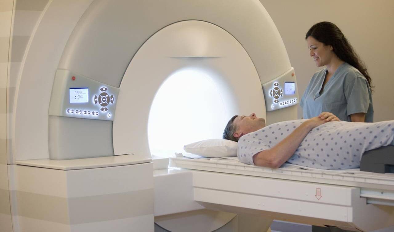
Magnetic Resonance Imaging (MRI) Principles, and Applications
Magnetic Resonance Imaging (MRI) is a sophisticated medical imaging technique that uses powerful magnets and radio waves to generate detailed images of the internal structures of the human body. Since its introduction in the 1970s, MRI has become an invaluable tool in medical diagnosis, offering non-invasive and high-resolution images for a wide range of clinical applications. This essay provides an overview of the principles behind MRI, its applications in medicine, and recent advances in the field.
Principles of Magnetic Resonance Imaging:
A. Magnetic Fields:
- MRI relies on the interaction of protons (hydrogen nuclei) within the body with strong magnetic fields.
- The primary magnet creates a static magnetic field, typically measured in Tesla (T), which aligns the protons within the body.
B. Radiofrequency Pulses:
- Short bursts of radiofrequency pulses are applied to the body, temporarily disturbing the alignment of protons.
- When the pulses are turned off, protons release energy and return to their aligned state, emitting radiofrequency signals.
C. Signal Detection:
- Radiofrequency signals are detected by receiving coils, and the data is processed by a computer to create detailed images.
- Variations in tissue composition and density influence the strength and timing of the signals, contributing to image contrast.
Clinical Applications of MRI:
A. Neuroimaging:
- MRI is widely used for imaging the brain and spinal cord.
- Applications include detecting tumors, assessing vascular abnormalities, and diagnosing neurological disorders such as multiple sclerosis.
B. Musculoskeletal Imaging:
- MRI provides detailed images of bones, joints, and soft tissues.
- It is valuable for evaluating injuries, detecting arthritis, and diagnosing conditions affecting muscles and tendons.
C. Abdominal and Pelvic Imaging:
- MRI is employed for visualizing abdominal organs, such as the liver, kidneys, and pancreas.
- Applications include detecting tumors, assessing organ function, and evaluating gastrointestinal conditions.
D. Cardiovascular Imaging:
- Cardiac MRI allows for the assessment of heart structure, function, and blood flow.
- It is utilized in diagnosing heart diseases, evaluating congenital abnormalities, and planning cardiac interventions.
E. Breast Imaging:
- Breast MRI is used as a supplemental imaging tool in breast cancer diagnosis.
- It provides additional information about tumor size, extent, and characteristics.
F. Functional MRI (fMRI):
- fMRI measures changes in blood flow and oxygenation to assess brain activity.
- Widely used in research and clinical settings to study brain function, map neural networks, and evaluate cognitive tasks.
Advanced MRI Techniques:
A. Diffusion-Weighted Imaging (DWI):
- Measures the random motion of water molecules in tissues.
- Applied in neurological imaging to detect acute stroke, tumors, and assess tissue microstructure.
B. Magnetic Resonance Spectroscopy (MRS):
- Provides chemical information about tissue composition.
- Applied in brain imaging to assess metabolites and detect abnormalities in conditions like brain tumors.
C. Magnetic Resonance Angiography (MRA):
- Visualizes blood vessels without the need for contrast agents.
- Used to assess vascular anatomy, detect aneurysms, and plan interventions.
D. Susceptibility-Weighted Imaging (SWI):
- Emphasizes variations in magnetic susceptibility.
- Valuable for detecting hemorrhages, vascular malformations, and certain brain disorders.
E. Functional Connectivity MRI (fcMRI):
- Maps functional connections between different brain regions.
- Applied in neuroscience research to study brain networks and understand disorders like schizophrenia.
ISafety Considerations and Contrasts:
A. Safety:
- MRI is considered safe with no ionizing radiation.
- However, precautions are necessary, such as screening for metal implants, as strong magnetic fields can interfere with metallic objects.
B. Contrast Agents:
- Gadolinium-based contrast agents may be used to enhance images, particularly in vascular and musculoskeletal imaging.
- Recent concerns about gadolinium retention in the body have led to increased scrutiny and cautious use.
Future Developments:
A. Artificial Intelligence (AI):
- AI applications in MRI include image reconstruction, automated image analysis, and improving diagnostic accuracy.
- AI algorithms may enhance efficiency and interpretation in clinical practice.
B. Ultra-High Field MRI:
- Increasing the magnetic field strength beyond 3 Tesla for improved spatial resolution and enhanced tissue characterization.
- Potential applications in neuroimaging, musculoskeletal imaging, and functional imaging.
C. Molecular Imaging:
- Developing MRI techniques to visualize specific molecules or cellular processes.
- Potential applications in cancer imaging, detecting inflammation, and monitoring treatment response.
Share this article

Magnetic Resonance Imaging (MRI) is a sophisticated medical imaging technique that uses powerful magnets and radio waves to generate detailed images of the internal structures of the human body. Since its introduction in the 1970s, MRI has become an invaluable tool in medical diagnosis, offering non-invasive and high-resolution images for a wide range of clinical applications. This essay provides an overview of the principles behind MRI, its applications in medicine, and recent advances in the field.
Principles of Magnetic Resonance Imaging:
A. Magnetic Fields:
- MRI relies on the interaction of protons (hydrogen nuclei) within the body with strong magnetic fields.
- The primary magnet creates a static magnetic field, typically measured in Tesla (T), which aligns the protons within the body.
B. Radiofrequency Pulses:
- Short bursts of radiofrequency pulses are applied to the body, temporarily disturbing the alignment of protons.
- When the pulses are turned off, protons release energy and return to their aligned state, emitting radiofrequency signals.
C. Signal Detection:
- Radiofrequency signals are detected by receiving coils, and the data is processed by a computer to create detailed images.
- Variations in tissue composition and density influence the strength and timing of the signals, contributing to image contrast.
Clinical Applications of MRI:
A. Neuroimaging:
- MRI is widely used for imaging the brain and spinal cord.
- Applications include detecting tumors, assessing vascular abnormalities, and diagnosing neurological disorders such as multiple sclerosis.
B. Musculoskeletal Imaging:
- MRI provides detailed images of bones, joints, and soft tissues.
- It is valuable for evaluating injuries, detecting arthritis, and diagnosing conditions affecting muscles and tendons.
C. Abdominal and Pelvic Imaging:
- MRI is employed for visualizing abdominal organs, such as the liver, kidneys, and pancreas.
- Applications include detecting tumors, assessing organ function, and evaluating gastrointestinal conditions.
D. Cardiovascular Imaging:
- Cardiac MRI allows for the assessment of heart structure, function, and blood flow.
- It is utilized in diagnosing heart diseases, evaluating congenital abnormalities, and planning cardiac interventions.
E. Breast Imaging:
- Breast MRI is used as a supplemental imaging tool in breast cancer diagnosis.
- It provides additional information about tumor size, extent, and characteristics.
F. Functional MRI (fMRI):
- fMRI measures changes in blood flow and oxygenation to assess brain activity.
- Widely used in research and clinical settings to study brain function, map neural networks, and evaluate cognitive tasks.
Advanced MRI Techniques:
A. Diffusion-Weighted Imaging (DWI):
- Measures the random motion of water molecules in tissues.
- Applied in neurological imaging to detect acute stroke, tumors, and assess tissue microstructure.
B. Magnetic Resonance Spectroscopy (MRS):
- Provides chemical information about tissue composition.
- Applied in brain imaging to assess metabolites and detect abnormalities in conditions like brain tumors.
C. Magnetic Resonance Angiography (MRA):
- Visualizes blood vessels without the need for contrast agents.
- Used to assess vascular anatomy, detect aneurysms, and plan interventions.
D. Susceptibility-Weighted Imaging (SWI):
- Emphasizes variations in magnetic susceptibility.
- Valuable for detecting hemorrhages, vascular malformations, and certain brain disorders.
E. Functional Connectivity MRI (fcMRI):
- Maps functional connections between different brain regions.
- Applied in neuroscience research to study brain networks and understand disorders like schizophrenia.
ISafety Considerations and Contrasts:
A. Safety:
- MRI is considered safe with no ionizing radiation.
- However, precautions are necessary, such as screening for metal implants, as strong magnetic fields can interfere with metallic objects.
B. Contrast Agents:
- Gadolinium-based contrast agents may be used to enhance images, particularly in vascular and musculoskeletal imaging.
- Recent concerns about gadolinium retention in the body have led to increased scrutiny and cautious use.
Future Developments:
A. Artificial Intelligence (AI):
- AI applications in MRI include image reconstruction, automated image analysis, and improving diagnostic accuracy.
- AI algorithms may enhance efficiency and interpretation in clinical practice.
B. Ultra-High Field MRI:
- Increasing the magnetic field strength beyond 3 Tesla for improved spatial resolution and enhanced tissue characterization.
- Potential applications in neuroimaging, musculoskeletal imaging, and functional imaging.
C. Molecular Imaging:
- Developing MRI techniques to visualize specific molecules or cellular processes.
- Potential applications in cancer imaging, detecting inflammation, and monitoring treatment response.
